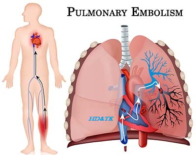
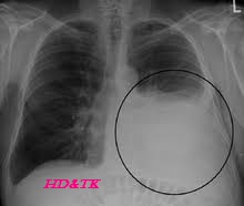
Solitary Pulmonary Nodule
A solitary pulmonary nodule is defined as a discrete, well-marginated, rounded opacity less than or equal to 3 cm in diameter that is completely surrounded by lung parenchyma, does not touch the hilum or mediastinum, and is not associated with adenopathy, atelectasis, or pleural effusion.
Differential Diagnosis
Malignant
bronchogenic, carcinoid, meta static cancer
Benign
healed infectious granuloma, benign tumors (hamartoma), AVM, rheumatoid nodule,
Wegener’s granulomatosis, hydatid cyst, round atelec tasis, intra pulmonary
lymph nodes, pseudotumor
Clinical Features
History
dyspnea, cough, hemoptysis, wheezing, chest pain, weight loss, fever, night
sweats, rheumatologic screen, past travel history, occupational expo sures,
medical history (smoking, lung cancer or other malignancies, TB, infections,
rheumatoid arthritis), medications
Physical
vitals, weight loss, clubbing, cyanosis, Horner’s syndrome, SVC syndrome,
lymphadenopathy, respiratory examination, abdominal examination (hepatomegaly),
bony tenderness
Investigations
Basic
Labs
CBCD, electrolytes, urea, Cr, LDH, AST, ALT, ALP, bilirubin, INR, PTT
Imaging
CXR, CT chest
Special
ABG
Screening
for Inflammatory Disorders ESR, CRP, ANA, ANCA
Biopsy
bronchoscopy or CT guided
PET/CT
SCAN if moderate to high suspicion of lung cancer

Diagnostic Issues
Findings
suggestive of Malignancy
Age
>50
Border
irregular, nodular cavity with thick wall, or speculation
Calcification
eccentric or uncalcified
Diameter
> 3 cm {> 1.2 in.]. If < 3 cm, 20 50% malignant. If > 3 cm,
50% malignant
Timings
if malignant, usually able to detect an increase in size of SPN between 30 days
and 2 years. Unlikely to be malignant if significant change in
Calcification Clues
Malignancy
eccentric/uncalcified calcification
Tuberculosis
or Histoplasmosis central/com platelets calcification
Benign
Hamartoma popcorn calcification
Management
Treatment
if low probability, observation with serial CT scans. If medium prob ability, bronchoscopy
with biopsy/brush or trans thoracic (CT/US guided) biopsy. If high probability,
thoracotomy with resection or video assisted thora coscopy (for patients who
cannot tolerate thoracotomy medically and physiologically)
Specific Entities
Pancoast
Tumor
Pathophysiology
superior sulcus tumors (mostly squamous cell carcinoma) invading and compres
sing the paravertebral sympathetic chain and brachial plexus
Clinical Features shoulder and arm pain (C8, T1, T2 distribution), Horner’s syndrome
(upper lidptosis, lower lid inverse ptosis, miosis, anhydrosis, enophthalmos,
absence of ciliary spinal reflex and heterochromia), and neurological symptoms
in the arm (intrinsic muscles weakness and atrophy, pain and paresthesia of 4th
and 5th digit). Other asso ciated findings include clubbing, lymphadenopa thy,
phrenic or recurrent laryngeal nerve palsy, and superior vena cava syndrome
Diagnosis
CXR, CT chest, percutaneous core biopsy
Treatments
concurrent chemoradiotherapy
Thoracic
Outlet Obstruction
Pathophysiology
Obstruction of the neurovascular bundle supplying the arm at the superior aperture
of the thorax. Common structures affected include the brachial plexus (C8/T1
> C5/C6/C7, 95%), subclavian vein (4%), and subclavian artery (1%)
CAUSES
anatomic (cervical ribs, congenital bands, subclavicular artery aneurysm),
repetitive hyperabduction/trauma (hyperextension injury, painters, musicians),
neoplasm (supraclavicular lymphadenopathy)
Clinical
Features triad of numbness, swelling and weakness of the affected upper limb,
particularly when carrying heavy objects. Brittle finger nails, Raynaud’s,
thenar wasting and weakness, sensory loss, decreased radial and brachial
pulses, pallor of limb with elevation, upper limb atrophy, drooping shoulders, supraclavicular
and infraclavi cular lymphadenopathy. Specific maneuvers include Roos test
(repeatedly clench and unclench fists with arms abducted and externally
rotated), modified Adson’s maneuver (Valsalva maneuver with the neck fully
extended, affected arm elevated, and the chin turned away from the involved
side), costoclavicular maneuver (shoulders thrust back ward and downward),
hyperabduction maneuver (raise hands above head with elbows flexed and
extending out laterally from the body), and Tinel’s maneuver (light percussion
of brachial plexus in supraclavicular fossa reproduces symptoms)
Diagnosis
cervical spine films, CXR, MRI
Treatments
conservative (keep arms down at night, avoiding hyperabduction), surgery

Comments
Post a Comment