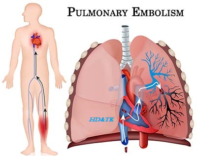
Human Histology
Histology
and its Study:
Histology
is the study of cell, tissues and organs as seen with a microscope.
Traditional
Histological Methods:
The
earliest histological observations were made on unfixed tissue (usually teased
make a flat preparation). The first advance was the discovery of chemicals for
fixation and staining of tissues. The next major development was invention of
instruments (called microtome) for cutting thin sections of tissue. These
sections could me mounted on glass slides and stained.
The
process of function preserves a tissue by denaturing its proteins. It also
makes the handling to tissue, and the preparation and staining of sections,
more efficient. Numerous fixatives are knows, the most commonly used being
formaldehyde (Formaldehyde is a gas, this gas dissolved in water is called
formalin).
Before
a tissue can be sectioned it has to be given a firm consistency, one way of doing
this to freeze the tissue and cut sections while it is still frozen (such
sections being called frozen sections).

Techniques
for the production of frozen sections have undergone great refinement and at
present they are prepared using a microtome enclosed in a refinement is called
cryostat.
Preparation
of frozen sections is the fastest method of examining a tissue. The technique
allows the examination of pieces of tissue removed by a surgeon, while the
patient is still on the operating table, making it possible for the surgeon to
plan his operation his keeping in mind the nature of disease.

Apart
from freezing a tissue, it can be made suitable for sectioning by the embedding
it in a suitable medium, the most common being paraffin wax. Such paraffin sections
can be thinner than frozen section and reveal more details of structure.
However some materials (e.g., fat) or last during the process of embedding
tissues in paraffin wax.
The
commonest staining procedure used in histology is baemataxylin-eosin staining.
In sections stained with most other components are seen in verging shades of
pink. Numerous other staining methods are available for demonstrating specific
tissue elements.
Electron microscopy:
In
the last few decades many new discoveries in field of histology have become
possible because of the development of the electron microscope (usually
abbreviated to EM). This microscope uses an electric fields in place of lenses.
With the EM magnifications in excess of 100,000 times can be achieved. The structure
of a cell or tissue as seen with the EM is referred to a ultra structure.

For
EM studies small pieces of tissues are fixed very rapidly after removal from
the animal body. Special fixatives are required (the most common being
glutaraldehyde). Very thin sections are required, and for this purpose thin
tissues have to be embedded in media that are harder than wax. The microtome
used for cuttings sections are such more sophisticated versions of traditional
microtome’s and are called ultramicrotomes. Thin sections prepared in this way
are also very useful in light microscopy. This reveal much more detail than can
be seen in conventional paraffin sections.
Before
sections are examined under an electron microscopy they are often treated with
solution containing uranium or lead, to increased contrast of the image. Osmium
tetraoxide acts both as fixative and staining agent and has been extensively
used for preparing tissues for electron-microscopy.
In
conventional EM studies (or transmission electron-microscopy) images are formed
by electron passing through the section. Wide use is also made of scanning
electron-microcopy in which the surface of tissue can be seen, and three
dimensional images can also be obtained. Especially useful details of some
tissues (e.g., membranes) can be obtained by freezing a tissue and then
fracturing it to view the fractured surface.
Histochemistry:
In
many cases the chemical nature of cellular and intra-cellular constituents can
be determined by the use of staining techniques. Lipids and carbohydrates
(glycogen) present in the cells are easily demonstrated. The presence of many
enzymes can be determined by placing section in solutions containing the
substrate of the enzyme, and by observing the product formed by action of
enzyme on substrate. The product is sometimes visible, or can be made visible
using appropriate staining agents.
For
enzyme studies the use of frozen sections in essential. Good frozen sections
can be obtained by using cryostats 9mentioned above).
Immunocytochemistry:
Specific
molecules within cells can be identified by treating tissue sections with
antibodies specific to the molecules. The technique enables chemical substances
o be localized in cells with great precision. Such studies have greatly
enhanced our knowledge of chemical transformations taking place within the
cells.
Autoradiography:
Many
molecules (e.g. aminoacids) injected into the animal become incorporated into
the tissues of the animal. Sometimes it is possible to replace a normal
aminoacid with a radioactive substitute. For example if a radioactive isotope
of thymidine. The sites of pressence of the radioactive material can be
determined by covering tissue sections with a photographic emulsion. Radiations
emerging frm radioactive material act on the emulsion.
Units
of measurement in Histology:
The
study of histology frequently involves the measurement of microscope distances.
The units used for this purpose are as follows.
1
micrometer or micron (um) = 1/1000 of a millimeter (mm).
1
nanometer (nm) = 1/1000 of a micrometer.
Cells,
Tissue And Organs:
The
human body, like that of most other animals and plants, is made up of units
called cells. Cells can differ greatly in their structure.

Comments
Post a Comment